Last week, I was giving my dog, Otis, a good ol’ head scratch. You know the kind: ruffling his fur, rubbing his ears, and holding him gently around his jowls while rubbing his face and neck.
He yelped in pain.
Of course, I investigated. I found a swollen bump on the side of his neck directly under the jaw bone. It didn’t look like a pustule and it didn’t have a wound or a bite. It was deep under the skin. I wondered if it was a swollen lymph node or an abscess. I’d never heard of salivary mucoceles.
The vet office was closed and my dog was not in any distress. I made a mental note to call my veterinarian the next morning.
Overnight, I bumped him by accident and he yelped in pain. Yep. We were going to the vet. The holiday weekend approaching, I was going to be proactive in the event this thing turned into an emergency.
I dropped him off (this was a work-in appointment) and I received a call from the vet a couple of hours later.
Dr. B recalled her exam and findings and said that she’d done a needle aspirate of the firm mass. She’d put together some slides to send off to pathology to identify all the cells she saw under the microscope. She listed all the diagnostic possibilities: Otis could have anything from an abscess to lymphoma. It would take several days for the pathology report to come back.
YIKES. That was a REALLY long holiday weekend.
So, I took to Google and looked up all of the vet’s listed possibilities. Hey, Google isn’t a vet, but it kept my mind occupied. As I searched, I found articles about salivary mucoceles. Images of dogs with salivary mucoceles looked just like Otis. I was convinced this is what Otis had!
Salivary Mucocele - What's that?
From the American College of Veterinary Surgeons website:
A salivary mucocele is a collection of saliva that’s leaked from a damaged salivary gland or duct and has accumulated in the tissue. It can be painful at first but then becomes a painless swelling in the neck or even in the mouth. The swelling fluctuates, but never completely goes away.
With Otis, the mass was a little bit smaller in the mornings, but by the evening, it was bigger. As previously mentioned, Otis’s mass was painful at first, but then was not painful.
4 types
- Cervical Mucocele: the most common type. Located in the neck, under the jaw, or in between the jaw.
- Sublingual Mucocele, also called a ranula: another common location for the location of a mucocele is on the floor of the mouth alongside the tongue.
- Pharyngeal Mucocele: this type is less common. It’s considered a variation of a cervical mucocele, but the fluid accumulation is found almost entirely within the throat.
- Zygomatic Mucocele: a very rare type of mucocele where the saliva is originating from the zygomatic salivary glands, located just below the eye.
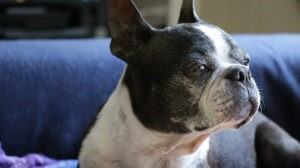
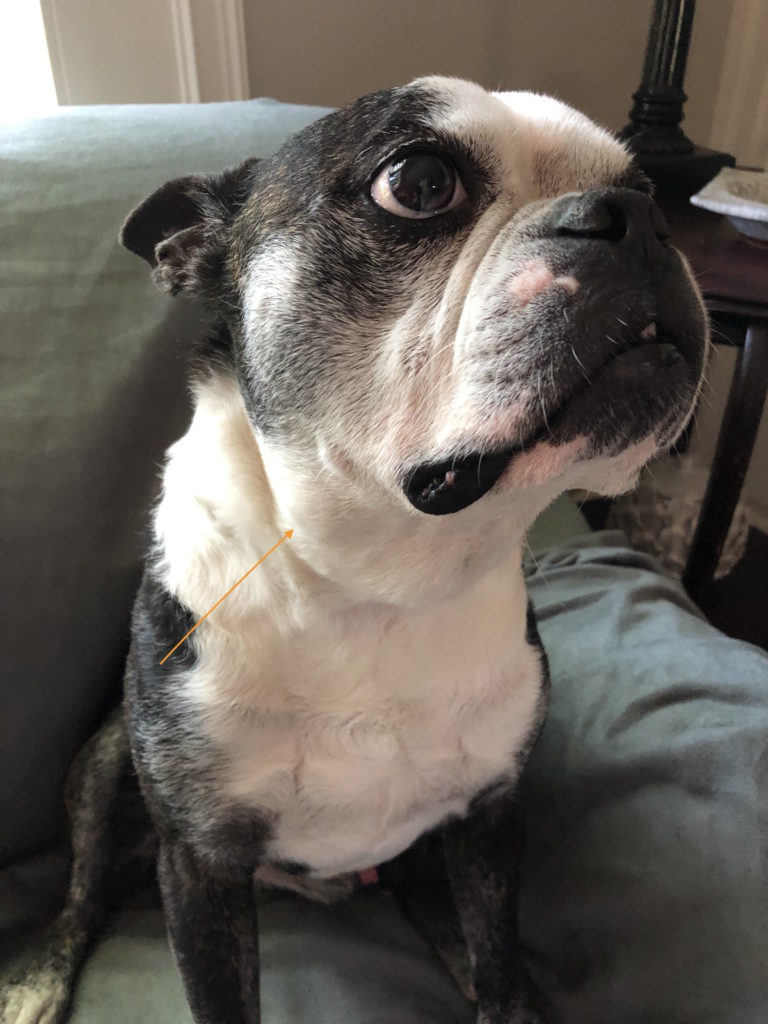
Otis’s neck looks like a cervical mucocele. It is under the lower jaw bone on his right side. Here’s what it looks like:
What causes a salivary mucocele?
A direct cause is rarely identified, but trauma is generally thought to be the most likely event. Choke collars, a bite to the face, or chewing on foreign materials is generally considered to be the most likely initiating event.
As the saliva leaks from the torn salivary gland or duct, it accumulates in the tissue and initiates an intense inflammatory response. A connective tissue capsule gradually forms around the saliva to prevent it from migrating elsewhere.
I have no idea how Otis could damage his salivary gland/duct. I don’t use choke collars. Heck, I don’t even use a martingale collar or slip lead on him. He hasn’t been in a fight. On occasion, he’ll chew a stick, but not often, and not for long, but I suppose that could be what happened.
Signs of a Salivary Mucocele
In the most common type of salivary mucocele, the cervical mucocele, the development is gradual and there are no real problems associated with the development of the mass. (That doesn’t mean you can leave it there.) With other types of mucoceles, there can be interference with eating or even breathing. With pharyngeal mucocele, sometimes the only sign is respiratory distress or difficulty swallowing as these mucoceles are undetectable except when a vet looks in the oral cavity. This requires sedation. Respiratory distress should never be ignored; treatment should be swift.
Otis doesn’t seem to be having any difficulty swallowing, although I’ve learned to not mess with his swollen area too much as it causes him to cough.
Salivary Mucocele - How Is The Diagnosis Confirmed?
According to the American College of Veterinary Surgery, the diagnosis of a salivary mucocele for most types is pretty straight forward. A veterinarian palpates the salivary gland. While tumors and abscesses may appear similar, these are generally painful on palpation and a salivary mucocele is not.
Otis’s was painful when I found this swollen mass, but by the time we arrived at the veterinary office, Otis did not express pain when the vet palpated his neck.
“If uncertainty exists about whether the mass is a mucocele or an abscess, a sterile aspiration of the fluid can be performed. Sometimes, a special laboratory testing (staining) can help confirm the type of fluid if there is doubt.”
Because the vet had some doubts about some of the cells she saw on her slides, we opted to send the slides to a laboratory for staining. The vet felt the swelling was a single lymph node, but because other findings were atypical, she wanted the help from a pathologist.
Treatment Recommendations for Salivary Mucocele
I noticed that Otis’s mass was visibly smaller for a couple of days following his vet visit and aspiration. This did not last long. His mass is just as big or slightly bigger than it was the day I found it.
Who Does These Surgeries?
Because of where the salivary mucoceles are generally located (remember, the cervical mucocele is located in the neck) surgical removal is considered a pretty operation. There are many important nerves and vessels in the neck. I know a couple of great veterinarians who are skilled at the surgery table, but those same veterinarians would refer me to a surgeon rather than do this surgery themselves.
If his diagnosis is confirmed to be a salivary mucocele, I will select a board certified surgeon to perform the surgery. I’m grateful my surgeon (who did three specialty surgeries on one of my dogs) is still in practice and accessible. I also have a backup surgeon I can choose if needed.
And The Diagnosis Is.......
NOT a Salivary Mucocele
See. This is why you don’t use Google to diagnose your dog. This is also why you pay for diagnostic testing.
Otis's Pathology Report Says.....
A pathology report can be a challenge for a non-medical person like me to interpret, (there’s a bunch of medical terms in the report!) but the report says this:
Microscopic Interpretation:
Consistent with a reactive lymph node.
Comments:
The cytologic findings are consistent with a reactive lymph node. There is no evidence of lymphoma, metastatic neoplasia, or lymphadenitis, lymph node appears to be due to lymphoid hyperplasia – a reactive lymph node. Causes for a reactive lymph node include infection, inflammation, immune-mediated disease or neoplasia from an area that drains into the node. No atypical cells or infectious organisms are seen.
What I understand the pathology report to say....
So – what I understand is this:
- A reactive lymph node is a normal way a dog’s body may respond to infection or inflammation or immune-mediated disease.
- It may signal something more severe than illness or infection.
The Treatment Plan
Our first approach is to use antibiotics. We chose to start with doxycycline. He’s been taking this medication for a few days. If there’s any reduction in the mass or improvement in the area, I can’t really see it, although I’m not an expert in feeling masses and measuring them. We have a second antibiotic to try, clindamycin, if we decide doxycycline is not working.
If, after a good trial of antibiotics, the lymph node is not improving, we will proceed with a tissue biopsy of the lymph node. A biopsy is much different than a needle aspirate. While a needle aspirate is less invasive, quick, and cost-effective, it’s a thin needle. It can be inserted into an area of a mass and miss. Meaning, important cells may not be in the exact location the needle is inserted.
A tissue biopsy is done in a couple of ways: a needle with a larger gauge may be used to extract tubular shapes of tissue from the mass. This is called a core biopsy or a punch biopsy. A biopsy can also be a resection of a mass, taking a chunk, or in the case of Rambo, the whole area is removed and sent off for evaluation by a pathologist. If I ever had any doubt about the value of using a pathologist, all doubt was erased when Rambo’s pathology report came back. It’s a great story should you care to read it.
I’m trying not to worry, but I am worried. I don’t like it when I don’t have answers. I don’t like unknowns. I’m confident we are on the right treatment course, but waiting sure is hard.
For now, I’m staying off Google.